|
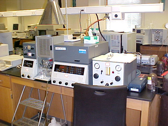
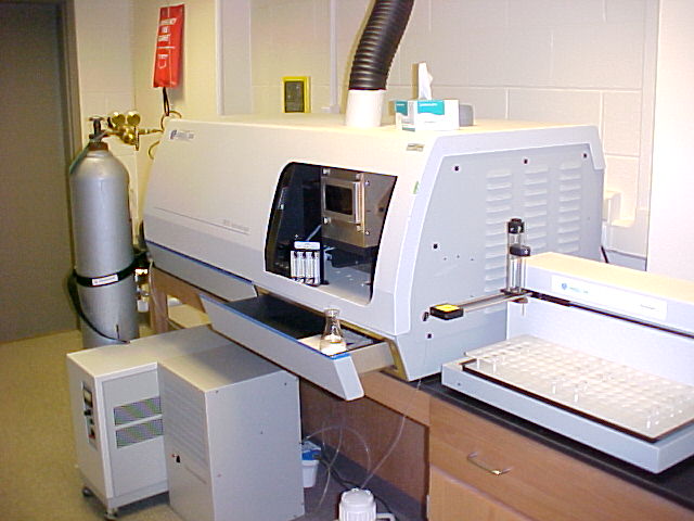
Flame Atomic
Absorption/Emission Spectrometer
Atomic spectrometers
are mostly used to measure metal concentrations in solution.
Solutions are aspirated into a flame, where thermal energy atomizes
the sample. The metal of interest emits or absorbs (from an element
specific lamp focused through the flame) specific wavelengths of light.
The radiant power absorbed or emitted at various concentrations allow
the determination of metals in complex samples such as lead in infant
blood, arsenic in soil, or manganese in water. |
|
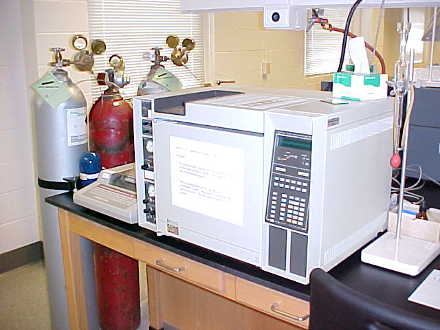
Gas Chromatograph
Gas chromatography is
typically used to separate, identify, and/or quantify volatile
compounds of a mixture. A carrier gas
passes through a 2-50 meter tube (column), which is either packed or
coated with specifically designed and chosen materials.
Solutions are volatilized by syringe-injection into a heated zone,
combined with carrier gas, and swept into the separation column.
Substances with strong affinity for the column material take longer to
pass through than those with weaker affinity, resulting in a separation
of the mixture. Programming the column's
oven temperature can enhance the separation.
Placing a detection device at the end of the column allows the
identification and quantification of the bands or peaks as they elute
from the column. Non-volatile or
thermally sensitive compounds are generally separated by liquid
chromatography. |
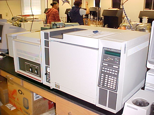 |
Gas Chromatograph-Mass
Spectrometer
A GC-MS system combines
the volatile compound separation technology of GC with the structure
determination power of mass spectrometry, resulting in a single
technique for separation, quantification, and identification of
components in a volatile mixture. GC
column effluents are typically fed into an ionization chamber and
bombarded with high energy electrons in order to ionize and fragment
the mixture components as each elutes.
Ions are directed to a detection system through a mass selector, which
is quickly scanned through a mass range producing a graphical plot of
ion intensity vs. mass/charge ratio, as well as a plot of total ion vs.
time. The total ion chromatogram peak area can be used to determine the
amount a component present, while mass spectra contains molecular and
fragment ion patterns that can help identify the substance.
The data collection workstation contains a mass spectral library of
over 50,000 compounds that can be used for comparison.
Drug testing at the Olympics is usually done by GC-MS. |
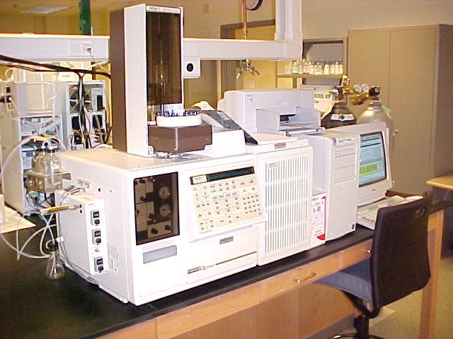 |
Gas Chromatograph-Ion
Trap Mass Spectrometer
As with other GC-MS
systems, an ion trap GC-MS system combines the volatile compound
separation technology of GC with the structure determination power of
mass spectrometry, resulting in a single technique for separation,
quantification, and identification of components in a volatile mixture.
Additionally, the ion trap allows the formation of gaseous anions and
cations that can be confined by the electric and/or magnetic fields for
an extended period of time. The trapping
and re-fragmenting of these ions can provide additional information
about the structure of the compound of interest. |
 |
Inductively Coupled
Plasma Atomic Emission Spectrometer
An inductively coupled
plasma atomic emission spectrometer is generally used to provide
qualitative and quantitative information about metals in solution
samples. Using an inert, inductively
heated, high temperature environment, efficient atomization and
spectral emission by the elements of interest is generally observed
when compared with other atomization techniques.
Combining the plasma with an echelle monochromator and two dimensional
array charge injection device detection system makes the determination
of many elements simultaneously possible. The axial orientation of the
plasma provides limits of detection generally an order of magnitude
lower than radially oriented plasmas.
The computer workstation's spectral library aids in the identification
of metals in unknown samples. |
|
Fourier Transform
Infrared Spectrometer
Infrared spectroscopy
is typically used to provide structural information about a substance
of interest. The substance is exposed to
a range of infrared frequencies resulting in a graphical plot of
radiant power absorbed (or transmitted) vs. frequency, wavelength, or
wavenumber. FT-IRs are equipped with a
Michelson interferometer in order to simultaneously measure all
infrared frequencies passing through a sample.
The resulting interferogram is deconvoluted by a Fourier analysis.
The resulting spectrum is a useful molecular fingerprint often
employed by forensic labs to analyze fibers, paint chips, or countless
other types of samples in solid, liquid,
or gaseous form. The data collection
workstation contains an infrared spectral library of over 50,000
compounds that can be used for comparison. |
 |
Polarimeter
Enantiomeric compounds
(optical isomers) are identical in almost all of their physical and
chemical properties. One notable
exception is the direction of rotation of the plane of vibration of
plane-polarized light. Plane polarized
light is light in which all wave vibrations have been filtered out
except for those in one plane. The
polarimeter is used to polarize light and then show the angle of
rotation of the plane of vibration by the optically active compound
placed in the light path. The amount of
rotation depends on the structure of the molecule, temperature
wavelength and concentration.
Polarimeters can be used to follow the course of reactions between
chiral compounds. |
|
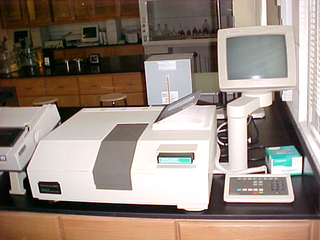
UV/Vis
Diode Array Spectrophotometer
UV/Vis spectrophotometry is typically used to provide quantitative or
structural information about a substance.
Samples are generally placed in a small cuvette and exposed to
ultraviolet and visible light. The
frequencies of unabsorbed light are dispersed and directed onto a
detection system constructed with a linear array of light sensitive
diodes. This array allows the
instantaneous measurement of the complete UV/Vis spectrum, rather than
monitoring a single frequency. The
computer workstation allows for timed events to be monitored.
An important use of diode array instruments
is to study enzyme kinetics. |
 |
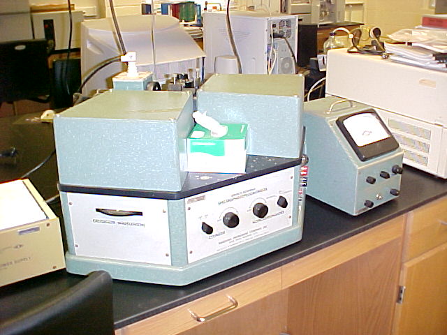
UV/Vis
Scanning Spectrophotometer
UV/Vis spectrophotometry is typically used to provide quantitative or
structural information about a substance.
Samples are generally placed in a small cuvette and exposed to
ultraviolet and/or visible light. The
frequencies of unabsorbed light are focused onto a photomultiplier tube
detection system using a double beam design.
Spectral bandwidths of 0.25 nm are possible.
Timed measurements are possible at fixed wavelengths for kinetic
studies. |
|
Spectrofluorometer
Fluorescence involves the absorption of light, from a light source, by
a substance, followed rapidly by an emission of light, at a lower
frequency, by the substance. Relatively
few substance exhibit this phenomenon making fluorescence a highly
selective technique. Fluorescence is
typically observed instrumentally at a right angle to the excitation
light source resulting in a highly sensitive technique.
Fluorescence is the detection technique commonly used in protein and
DNA sequencing, and in the detection of many environmental pollutants. |
|
NMR Spectrometer
Nuclear Magnetic
Resonance spectroscopy is probably the most important technique used
today for the structural study of organic and inorganic compounds.
Substances are placed into a large magnetic field (commonly a
superconducting magnet using a wire coil cooled to 4 K in a reservoir
of liquid helium). The spinning nucleus
of an atom either aligns with or against the externally applied
magnetic field. Radio frequency radiation
is used to "flip" the nuclear spin.
The frequency absorbed by a nucleus is determined by the local
electron density near each nucleus and by the geometry of the molecule.
NMR spectra are used to determine protein structure and for countless
other applications. |
|
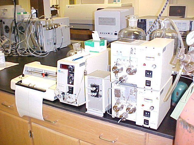
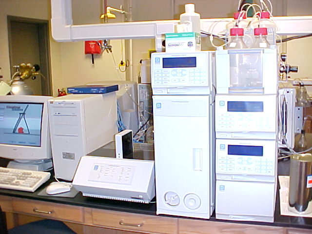
High Performance Liquid
Chromatograph
Liquid
chromatography is
typically used to separate, identify, and/or quantify compounds in a
mixture. A carrier solution (mobile
phase) is forced by means of a pump, through a 10-30 centimeter tube,
or column, which is packed with a specifically designed and chosen
material. Mixture solutions are
syringe-injected into the mobile phase and carried into the separation
column. Substances with strong affinity
for the packing material take longer to pass through than those with
weaker affinity, resulting in a separation of the mixture. The
mobile phase can be held in a fixed composition (isocratic mode) or
varied to enhance the separation by using multiple pumps and a
controller (gradient elution mode). Placing a detection device at the
end of the column allows the identification and quantification of the
bands or peaks as they elute from the column.
HPLC is an important technique for the
pharmaceutical industry, used to determine the purity of medications
and to quantify therapeutic components.
It is also widely used in the flavor and environmental industries.
Volatile mixtures are commonly analyzed by gas chromatography. |
|
Ion Chromatograph
Ion chromatography is a form of liquid chromatography in which the
substances of interest usually carry a formal charge.
The column packing material contains ionic sites, that have an
attraction for oppositely charged . These
sites serve as ion exchangers. Ions are
separated based on their charge and size. Ion chromatography is a
widely used technique for the analysis of ions in environmental samples. |
|
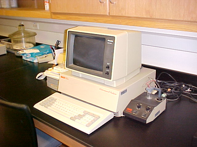
Electrochemical Analyzer (BAS100A)
Electrochemical analysis is a widely used technique for analyzing
substances in our bodies that can be easily reduced or oxidized.
Since many biochemical processes involve redox reactions, there are
many useful applications of these techniques.
Electrochemical analysis is also useful for the trace detection of
metals. |
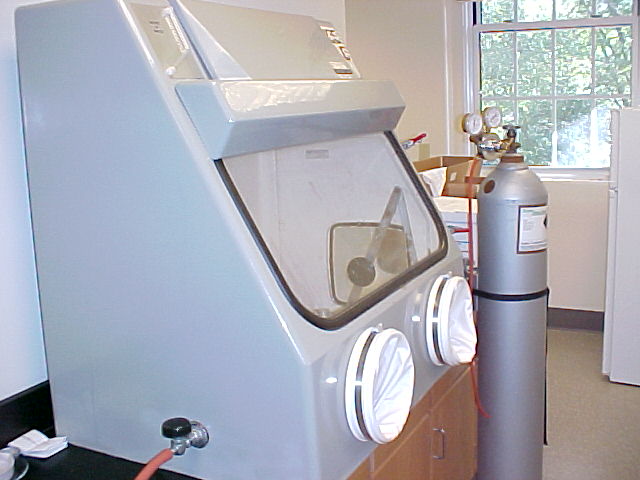 |
Inert Atmosphere Glove
Box
The inert atmosphere
glove box, or sometimes "dry box",
is used for the synthesis, storage and manipulation of air or moisture
sensitive materials. The workspace is typically filled with nitrogen,
argon or other inert gas providing a dry, air free environment.
Arm-length gloves and viewing window provide a means of working with
materials inside the chamber. An antechamber, which can be evacuated or
flooded with inert gas allows introducing materials or equipment from
the room into the inert atmosphere. Glove boxes have a central and
indispensable role in the study of air-sensitive compounds. |
|
Protein Perfusion
Chromatograph
Protein perfusion chromatography is an advanced technique for the rapid
separation of proteins. Samples pass
through small beads that have pores specially designed for protein
separation. Effluents are collected and
repurified. This instrument is used to
separate cell components to obtain pure protein samples for
crystallization and for structure determination with X-Ray diffraction. |